Cool newcomer
Biophysicist Emiliya Poghosyan grips a tiny object with a pair of tweezers and holds it up to the light. A closer look reveals it to be a circular copper lattice, only three millimetres in diameter. These specimen holders, known as grids, are essential tools for all scientists who, like Poghosyan, use electron microscopy for their research. "We deposit our sample on the small grids – although in the end the sample layer may be no more than 100 nanometres thick," Poghosyan explains. That is around five-hundredths the thickness of a human hair. Only in this way is it possible to capture and study individual molecules such as proteins under the microscope.
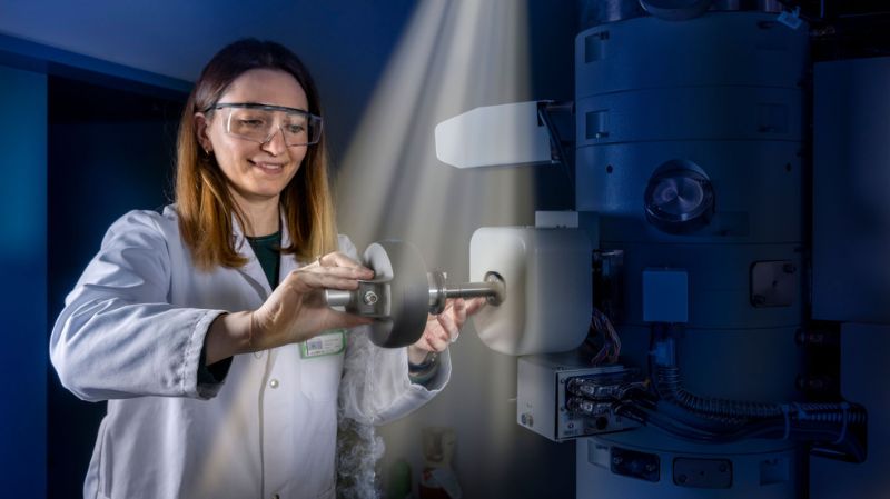
"It’s fascinating how rapidly electron microscopy has developed in recent years," says Emiliya Poghosyan, a scientist in the Laboratory for Nanoscale Biology, who is responsible for the electron microscopy instruments at PSI.
Basically, an electron microscope works like an ordinary light microscope; instead of using normal light, however, the objects to be examined are irradiated with electrons in a vacuum, thus achieving a resolution that is 2,000 times higher than with the best light microscope, allowing much smaller objects to be examined.
"The resolution is now so powerful that we can use this method to determine the three-dimensional structure of proteins and other biomolecules." The advances that have made this possible include a new generation of detectors that register electrons directly and analysis methods that compare the recordings of millions of molecules in a sample and, as it were, average them. This yields images with an improved signal-to-noise ratio, from which sharply defined molecular structures can then be determined. The so-called “resolution revolution” began less than ten years ago.
For biological samples, the breakthrough came with cryo-electron microscopy, the scientist explains. The samples are plunge-frozen before measurement. The cold protects the sensitive samples from damage that would inevitably be caused by the impact of the fast electrons. "Compared to measurements at room temperature, we can shoot around a hundred times as many electrons at the sample before it is destroyed," says Poghosyan. "And every additional electron increases the signal and the amount of information that we get during the measurement."
No ice crystals allowed
Emiliya Poghosyan clamps the tweezers with the copper grid into an elongated device, the vitrobot. She applies a tiny drop of her sample solution through a circular opening on the side of the apparatus. Two parts of the device that look like headphones then close around the grid. "They remove excess water from the sample," explains Poghosyan.
Then comes the temperature shock: the tweezers immerse the coated grid in a container with a liquid at a temperature of -196 degrees Celsius. This is ethane, cooled with liquid nitrogen. The sample freezes in a fraction of a second. "It is important for this to happen especially rapidly, because otherwise ice crystals can form and destroy the sample," the scientist explains.
Electrons cannot penetrate thick ice crystals, so such areas would later appear black in the image – this part of the picture is then ruined. If, on the other hand, water is cooled very quickly, it solidifies without crystallising. The result is vitreous water which, like a liquid, exhibits a disordered molecular structure and will be penetrated by the electron beams.
The goal is for the sample molecules to be evenly distributed in the holes in the grid, surrounded by a layer of glass-like ice that is as thin as possible. For this, the sample films may not be thicker than the biomolecules themselves. "The preparation of the sample grids is a science in itself," says the biophysicist. "There is no general-purpose recipe. You just have to keep trying new conditions until you have found the ideal one for a particular molecule."
The developers of cryo-electron microscopy, Swiss chemist Jacques Dubochet as well as Joachim Frank and Richard Henderson, were awarded the Nobel Prize in Chemistry in 2017 for their work. It was Dubochet who discovered the trick with the crystal-free water.
The ‘Aha’ effect comes later
With nitrogen cooling and a suitable amount of water vapour, Emiliya Poghosyan transfers her grid to the chilled mount of the electron microscope and pushes it into the larger than man-sized device. Then she diligently pours in liquid nitrogen. "You need patience when you work with cryo-electron microscopy," she says with a laugh. "If you don’t wait until all the equipment has cooled down sufficiently, the sample will be destroyed and all the work will have been in vain."
Finally, in the room next door, she can look at her sample on the monitor and take pictures. One individual image is not very impressive: basically, you only see a lot of small greyish spots on a light background. These spots are the protein molecules.
The ‘Aha’ effect comes with the subsequent data analysis: millions of molecules are averaged from a few thousand recordings. In this way, the protein is captured from all sides, since ideally a sample contains millions of molecules in many different orientations. Putting all this information together creates a three-dimensional model of the protein that is remarkably accurate. "It’s amazing how such precise 3D models can be created from these images," says Emiliya Poghosyan.
A path to success even without a crystal
"Electron microscopy has revolutionised the way we get structural information from proteins," says Jacopo Marino, a biologist in the PSI Laboratory for Biomolecular Research. "The whole process is advancing very rapidly."
A few years ago, a team at PSI including Jacopo Marino solved the structure of a complex consisting of the light receptor rhodopsin and a G protein – proteins in the retina that enable us to see. Knowing how the receptor docks with the G protein provides clues as to how signal transmission works in the cell and how this process can be manipulated under certain circumstances (see infographic page 16). With cryo-electron microscopy, the structure was solved within four months. And even that is still relatively long. "With proteins that are biochemically easy to manipulate, we can even have the structure in our hands within a few days."
The complex, which consists of several proteins is very flexible - a fact that stood in the way of X-ray crytallography. This flexibility means that obtaining crystals of the complex, essential for X-ray crystallography is extremely difficult, and couldn’t be obtained even with years of effort.
Currently, it is not possible to examine very small biomolecules with cryo-electron microscopy, says Emiliya Poghosyan. For instance, the structure of the protein rhodopsin alone cannot be deciphered in this way. In addition, the resolution is still marginally better in X-ray crystallography.
Complement, not replacement
Cryo-electron microscopy has enabled enormous progress in the investigation of membrane proteins such as receptors, which are naturally embedded in a cell membrane and are difficult to isolate in a pure form, let alone to make or to crystallise. One of the first proteins that Nobel Prize winner Richard Henderson worked with was bacteriorhodopsin, a membrane protein found in the cell wall of certain bacteria. Currently, Jacopo Marino is using this method to take a close look at an ion channel that plays a major role in signal transmission during the visual process.
Even very small amounts of protein are sufficient for cryo-electron microscopy. This makes the work easier and shortens the time it takes, especially with molecules that have to be laboriously isolated from tissues and cells. Also, the researchers don’t have to ensure their samples are meticulously clean.
However, cryo-electron microscopy will not replace the X-ray crystallography at SLS and SwissFEL. "The two are not in competition with each other," emphasises Marino. "They complement each other. Both have their strengths, and their limitations."
