Watching and analyzing T cells attack cancer cells in real time
Immuno-oncology is a promising new field of research that involves boosting the capacity of a patient’s own immune system to attack cancer cells. It has already proven to be effective in treating certain types of cancer, and scientists are now testing an array of molecules to expand the range of potential applications. These tests require a variety of instruments, each of which, perform a specific analysis. However, the measurements taken by these instruments are often limited and only provide an indirect indication of how immune cells interact with cancer cells. To enable scientists to quantify these interactions directly, Nanolive has developed a new test, called the Live T Cell Assay, that lets scientists observe T cell behavior in real time and generate quantitative analysis of numerous cell characteristics. The Live T Cell Assay is intended to be a turnkey solution for evaluating next-generation immunotherapy drug candidates.
T cells as serial killers
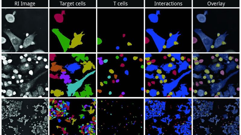
Nanolive’s offering consists of a unique automated holotomographic microscope based on a revolutionary technology developed by Nanolive’s CEO Yann Cotte during his PhD studies at EPFL a few years ago, along with algorithms that have been trained to recognize and extract data on T cells and the cancer cells they target. It employs imaging methods that leave the cells intact and allow scientists to observe them live over time. More specifically, the system uses holotomography to generate 3D images of the distribution of refractive indices of the cell samples, providing detailed data on the cells’ structures and chemical composition. By combining the holotomography with artificial intelligence and advanced computer vision techniques, it can distinguish T cells from cancer cells without the need for markers and track the cells’ interactions directly. “Our algorithms can sort through all these parameters and extract data on the different phases of the cells’ interaction, from the detection of binding and stress responses in cancer cells, to the moment they are eventually killed,” says Mathieu Frechin, the head of Artificial Intelligence for Quantitative Biology at Nanolive. “We’ve even seen that some T cells act as ruthless serial killers!”
Automated analyses

© 2021 Nanolive
Drug developers in the pharmaceutical industry need to be able to rapidly test many different drug candidates on cancer cells in order to identify which ones work best. That’s where Nanolive’s new system comes in: it allows scientists to test several drug candidates simultaneously under optimal experimental conditions, and extract automated analyses summarized in customizable charts and graphs.
What’s more, by allowing scientists to watch cell interactions live, the system also reveals phenomena that would be impossible to observe if the cells had to be immobilized. “This is the first time we are able to see so nicely the killing process of T cells involved in in vitro assays. We can also extract additional key parameters like the T cell killing rate, and the average number of cancer cells killed by one T cell, which is very important because these type of parameters are not easily extracted using conventional methods,” says Valery Moine, Head of Pharmacology Unit Light Chain Bioscience.
Improving clinical success rates
A further benefit of the Live T Cell Assay is that it can enhance downstream efficacy, improve the success rates of clinical trials and limit the need for in vivo testing. It does so by allowing scientists to fully characterize, in the very first phases of the drug discovery process, how specific chemical compounds affect the interaction between T cells and cancer cells. What’s more, the information revealed by the system’s imaging technology goes beyond the automated analyses, as experienced users can comb through the data it generates and run their own evaluations. Because Nanolive’s technique is non-invasive, the precious samples from patients can be reused for downstream applications such as cell therapy or personalized medicine.
