X-ray microscopy with 1000 tomograms per second
Most people are familiar with computed tomography from medicine: a part of the body is X-rayed from all sides and a three-dimensional image is then calculated, from which any sectional images can be created for diagnosis.
This method is also very useful for material analysis, non-destructive quality testing or in the development of new functional materials. However, to examine such materials with high spatial resolution and in the shortest possible time, the particularly intense X-ray light of a synchrotron light source is required. In the synchrotron light, even rapid changes and processes in material samples can be visualised if it is possible to capture 3-dimensional images in a very short time sequence.
A team led by Francisco García Moreno from the Helmholtz Centre Berlin is working on this, together with researchers from the Swiss Light Source SLS at the Paul Scherrer Institute (PSI). Two years ago, they managed a record 200 tomograms per second, calling the method of fast imaging “tomoscopy”. Now the team has achieved a new world record: with 1000 tomograms per second, they can now record even faster processes in materials or during the manufacturing process. This is achieved without any major compromises in the other parameters: the spatial resolution is still very good at several micrometres, the field of view is several square millimetres and continuous recording periods of up to several minutes are possible.
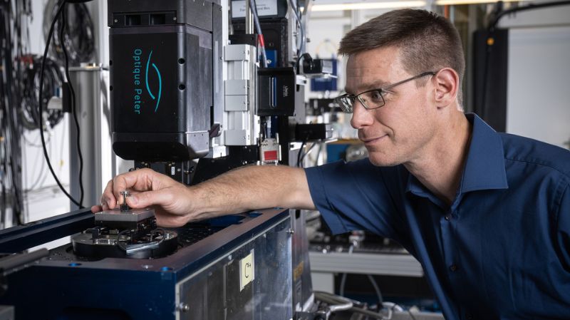
Special table reaches 500 rotations per second
For the X-ray images, the sample is placed on a high-speed rotary table developed in-house, whose angular speed can be perfectly synchronised with the camera's acquisition speed. "We used particularly lightweight components for this rotary table so that it can turn around its axis 500 times per second and still remain stable," García Moreno explains.
Creating a 3D image from 40 projections per millisecond
At the Tomcat beamline at the SLS, which is specialised in time-resolved X-ray imaging, PSI physicist Christian Schlepütz used a new high-speed camera and special optics. "This increases the sensitivity very significantly, so that we can take 40 2D projections in one millisecond, from which we create a tomogram," Schlepütz explains. One 3D image is therefore created every millisecond, in other words 1,000 3D images per second. With the planned SLS2.0 upgrade, even faster measurements with higher spatial resolution should be possible from 2025.
The team demonstrated the power of tomoscopy with various examples from materials research: the images show the extremely rapid changes during the burning of a sparkler, the formation of dendrites during the solidification of casting alloys or the growth and coalescence of bubbles in a liquid metal foam. Such metal foams based on aluminium alloys are being investigated as lightweight materials, for example for the construction of electric cars. The morphology, size and cross-linking of the bubbles are important to achieve the desired mechanical properties such as strength and stiffness in large components.
"This method opens a door for the non-destructive study of fast processes in materials, which is what many research groups and also industry have been waiting for," says García Moreno.
Text: based on a press release from the Helmholtz Centre Berlin with additions from the Paul Scherrer Institute
