X-ray imaging after heart transplantations
One of the biggest concerns following a heart transplant is the possibility of the patient’s immune system rejecting the new organ. Although immunosuppressants – drugs that suppress the immune system’s response – prevent organ rejection, they also weaken the body’s natural protection against pathogens, so balancing between the two perils is needed.
To establish whether levels of immunosuppressants are sufficient, tissue samples are usually taken from the transplanted heart during follow-up examinations. In the current system, these biopsies first have to be fixed, sliced, and stained before they can be examined under the optical microscope. If immune cells are found to have migrated into the tissue and caused damage, this indicates a rejection reaction and clinicians then need to decide whether the patient needs to be given additional immunosuppressants, depending on the severity of the rejection.
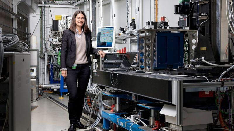
Now a study has shown that X-ray phase contrast imaging using synchrotron light is at least as effective as a standard optical microscope for screening the biopsy material. Moreover, by improving the diagnosis, the method could lead to less frequent and more targeted administration of immunosuppressants and thus reduce the dosage of those drugs. The study was conducted by a team of clinicians and biomedical scientists around Anne Bonnin, a researcher at the Laboratory for Macromolecules and Bioimaging. “We hope that in the future, this technique will improve diagnostics in follow-up after heart transplants,” says Bonnin.
3D images of endomyocardial tissue
As part of a small-scale initial study, researchers used the TOMCAT beamline of the Swiss Light Source SLS to analyse tissue samples taken from the hearts of 23 patients who had undergone a heart transplant at University Hospital Centre Zagreb. No time-consuming preparation or staining of the tissue samples is necessary: the samples are simply fixed in formalin and scanned for about twelve minutes virtually in their natural state. This produces high-resolution 3D images that can be manipulated on the computer screen as needed. Clinicians and pathologists can zoom in on the heart tissue to inspect details of less than one thousandth of a millimetre.
In this study, an experienced clinical pathologist screened these 3D images, as well as 2D slices taken from these images, and assessed the degree of rejection separately in the usual way. In parallel, an assessment was made using the classical histopathological method. The conclusion: the agreement between assessments was high, as confirmed by the statistics. Thanks to the 3D visualisation of one sample out of the 23, a misdiagnosed case by classical histopathology was found.
Due to its three-dimensional character, the new method could be superior to the established method, the researchers believe. “On the 3D images of the entire sample, for example, it’s possible to determine that the immune cells have only penetrated a small part of the sample, so the damage is not as bad as might appear on a selected 2D slice,” Anne Bonnin explains. “In this case, there is perhaps no need for escalation of immunosuppressants, so the patient is spared undesirable side effects and the health system saves on unnecessary costs for expensive drugs.”
A bigger follow-up study on heart transplants
A more ambitious follow-up study further exploring the method’s potential has already started, funded by the Croatian Science Foundation. In addition to University Hospital Centre Zagreb, University Hospital Dubrava, Zagreb (Croatia), and Hospital Clinic, Barcelona (Spain), are also involved and are recruiting heart transplant patients for the study.
Synchrotron light is particularly intensive X-ray light capable of visualising details that conventional X-rays cannot pick up. Phase contrast enhanced imaging using synchrotron light is especially suitable for biomedical applications. The PSI research group led by Marco Stampanoni, in which Anne Bonnin is working, have already used this method to examine, among other things, the vascular system of the mouse brain as well as lung tissue from rats during the respiratory process. They have also illuminated the heart of a human foetus to determine which abnormalities may have caused the premature death of an unborn child.
The technique could be extremely useful for diagnostics in hospitals. In the long term, hospitals should no longer need to send their biopsies for analysis to large-scale facilities such as the SLS. “In the near future, we hope to be able to develop a phase-contrast system that provides such type of 3D virtual histology images and compact enough to be installed in a hospital,” says Anne Bonnin. PSI researchers are already working toward this, by transferring the techniques developed at the SLS in a way that they can also be used with laboratory X-ray sources.
