Molecular atlas reveals how brain cells develop
As the fertilized egg divides, initially undifferentiated cells take on specific functions, becoming more distinct as different tissues and organs emerge. Understanding how hundreds of disparate cell types arise has proven difficult, largely because scientists have lacked the technologies to capture cellular decision making over time.
Recent advances have allowed researchers to measure changes in gene activity of individual cells, so several groups started to study in detail how specialized cell types are formed in specific brain regions. However, nobody had thus far traced the patterns of gene expression across the entire developing brain.
Now, for the first time, EPFL researchers and their collaborators at Karolinska Institute in Sweden mapped the genetic and developmental trajectories that embryonic cells follow toward their fate in the maturing brain. This molecular atlas could not only help better understand the healthy and diseased brain, but also improve therapeutic approaches such as cell replacement therapy for neurodegenerative diseases, says study lead author Gioele La Manno, head of the Laboratory of Neurodevelopmental Systems Biology at EPFL. The findings were published in Nature.
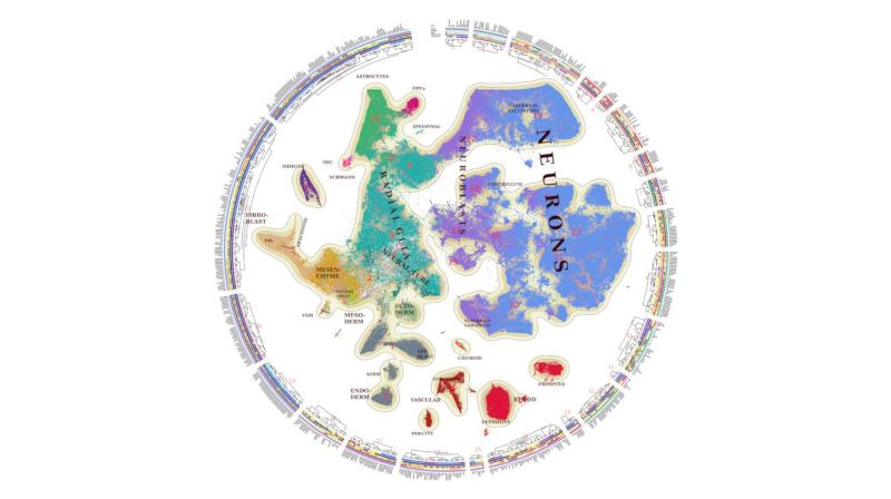
To monitor decision making in individual cells over time, La Manno and his colleagues analyzed brain samples from mouse embryos every day from day 7 after fertilization until birth. Using a combination of powerful sequencing techniques and mathematical methods, the researchers obtained about 290’000 gene expression profiles of individual cells from all brain regions, as well as nearly 800 cellular ‘states’ that included the developmental programs for different cells, including neurons and neuronal support cells.
As neuronal progenitors mature, they stop proliferating and differentiate into scores of different neurons. The researchers tracked the emergence of this diversity and described the timing of appearance of primitive nerve cells, called neuroblasts, across different brain regions. In mice, the first neuroblasts appear early, before day 9 of embryonic development — which roughly corresponds to the beginning of the first pregnancy trimester in humans. These pioneer neurons are involved in sensory and motor functions, the researchers found. “One of the first things to do is to set up the motor and sensory functions, because if you don’t set these up early, later on it will become more difficult to build ‘highways’ towards the periphery,” La Manno says.
The researchers also found specific types of neuronal progenitors, called organizer radial glial cells, whose role is to guide the development of neighboring cells by producing molecular messengers that help establish the position of various specialized cell types within the brain. “If the brain were an orchestra, organizer radial glial cells would be the director,” La Manno says. These radial glial cells produce a greater variety of molecular messengers than scientists thought, the team found.
The analysis also allowed the researchers to identify cell populations of different sizes, with some populations made of 100 times more cells than others. One of the biggest populations appears to be that of excitatory neurons in the forebrain, an area that comprises most regions involved in higher-order cognition. One of the smallest populations identified was that of a type of neuronal support cell called ependymal cells, which produce the fluid that surrounds the brain and spinal cord.
La Manno hopes that the wealth of information contained in this brain atlas could help identify genes involved in neurodevelopmental conditions and determine the origin of malignant cells in brain cancer. The atlas, he says, could also serve as a reference to evaluate brain tissues generated from stem cells in a laboratory dish. To help others study cells and tissues of medical interest, the researchers made the atlas publicly available as a browsable web resource.
Next, La Manno plans to uncover where in the developing brain the different cell populations are located. “The current atlas is a molecular chart that tells you which kind of cells are similar and which ones are different,” La Manno says. “Now, we want to see where these cells sit within the brain.”
