Watching receptor proteins changing shape
Mr. Guixà-González, you’ve been doing research on G protein-coupled receptors for many years. Why are they so interesting?
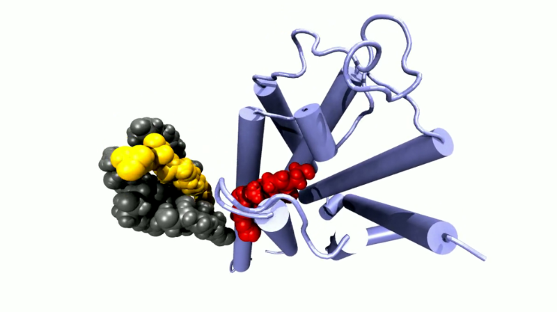
G protein-coupled receptors are just everywhere – almost every cell in our body needs them. These protein molecules protrude from a cell like antennas. Molecules that circulate in the bloodstream, for example, dock on them. They are therefore also important targets for active agents in medicine. In fact, it is estimated that more than one-third of all currently approved drugs owe their efficacy to this type of protein.
How, precisely, do these receptors act?
They are embedded in the cell membrane and convey information into the cell. Let's assume you're excited and the body has released adrenaline, which is now circulating in the blood. It then binds, for example, to the G protein-coupled receptor in the membrane of a heart muscle cell. Then the receptor changes its shape: It becomes activated, as we say. In its active state, the receptor binds proteins inside the cell. If the receptor now, under the influence of adrenaline, changes its shape, the intracellular protein is released and exerts some function. In the case of the heart muscle cell, an enzyme is activated that then, through a cascade, ultimately causes the heart to beat faster.
And presumably something different happens in other types of cells?
Right. There are over 800 different types of G protein-coupled receptors. Besides adrenaline, they can register loads of other things: hormones and neurotransmitters in the brain, for instance. Likewise for sight and taste. Also, if we smell something, molecules bind to such receptors.
How can drugs intervene in this process?
Let's assume someone has a disease in which one of the receptors is permanently activated. So you develop a drug that binds to the receptor and blocks it. Or, conversely, someone wants to develop a drug that activates the receptor. In both cases we need a molecule that fits to the receptor precisely.
Is that a bit like solving a puzzle where you look for two pieces that fit together?
Roughly, yes. However – unlike a puzzle – it’s not just about the static structure of the receptor. In reality there’s a whole movie playing. Because when a molecule binds to the receptor, it thereby changes its shape. And even without activation, the whole thing is a very dynamic system: Interactions within the protein are constantly being loosened and stabilised again. The question is: How, precisely, do these changes proceed?
So you want to see the whole movie and not just individual images. How do you go about doing this?
It's a bit like the early days of photography. Back then, people didn't know exactly how horses gallop: Do they have all four hooves on the ground at some point or not? You couldn't see it with the naked eye, because the horses were too fast. There weren't any video cameras yet. To observe the process, you can take a lot of photographs, lay them out one after the other and fill the gaps using computer simulations. The horse is our receptor protein, and the photographs are crystal structures.
Crystal structures?
Yes, in structural biology you often work with crystal structures: You determine the structure of a biomolecule by bombarding crystals of it with X-ray light and then calculating the three-dimensional structure from the resulting pattern. Such crystal structures are basically just snapshots. We take such crystal structures, which researchers have determined with the Swiss Light Source SLS or the X-ray free-electron laser SwissFEL, create a model on that basis, and then look at how the molecules move according to the laws of classical physics. That’s how we get our film.
How can the movements of the receptors be calculated?
Using the crystal structures, we generate a three-dimensional atomic model of the system. It shows the position of every single atom within the protein. In addition, the model contains the lipid molecules of the cell membrane as well as water molecules and salt ions. Such a model can easily contain 100,000 atoms or more. Using the technique of molecular dynamics, we then look at the interactions between individual atoms or parts of the protein: How large are the distances, the angles, and the energies, how strong are the interactions, and what spatial motions result from them? So the atoms virtually begin to dance – a very physical dance according to the rules of classical physics. With this method we simulate the movements of the protein over several microseconds. These calculations take several weeks, however, and we need supercomputers to carry them out.
So it requires a tremendous amount of computing power. But doesn't the research at SwissFEL take on this same task, that is, creating movies of molecular motion?
The idea behind an X-ray free-electron laser is as follows: Instead of taking a single picture of a static protein, you take several pictures at very short intervals while the protein is moving. If you put these images – that is, crystal structures – one after the other, you can actually see how the protein moves. But at the moment SwissFEL cannot replace the molecular dynamics method. That’s because so far only certain proteins – mostly those that are activated by light – can be examined in SwissFEL. Most proteins, however, are activated by other things, such as hormones or other proteins. In such cases, molecular dynamics has so far been the method best suited for studying protein movements at atomic-scale resolution. At least at this moment.
Together with a large European research consortium and under the direction of Jana Selent from Pompeu Fabra University at Barcelona, Spain, you launched the online platform gpcrmd.org – short for “G protein-coupled receptors molecular dynamics”. What was the goal?
In our research field, many of us create dynamic simulations of G protein-coupled receptors. With the new platform, we want to make as many of these simulations as possible generally accessible to science. Researchers from all over the world can now share their simulations with each other and analyse those of other groups. Up to now, this is unique.
How many simulations does the platform include so far?
More than 600 at this time. This means that we cover 70 percent of all families of G protein-coupled receptors. We are currently in the process of preparing a second version of the platform. This should then include at least one simulation for each family.
How does the platform help in drug development?
You can, for example, look at how different molecules bind to the receptor – and thus check how you might be able to improve your potential active agent. You can also watch how water molecules typically distribute themselves around the receptor and influence the effect of bound molecules. Or you can investigate from what directions an active substance is able to reach the receptor at all. The platform offers countless possibilities – and all for free!
Interview: Paul Scherrer Institute/Brigitte Osterath
