Imaging at the next level
Two researchers with a shared interest who want to examine the structure of samples that are several millimetres – or even centimetres – in size in three dimensions with a resolution of a few millionths of a millimetre and, above all, make them visible in imaging. Both have been awarded an ERC Starting Grant from the European Research Council, enabling them to establish and develop their own research area.
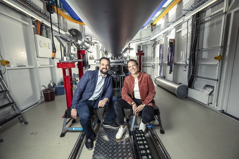
Marianne Liebi is an assistant professor at EPFL and a group leader at PSI who holds a doctorate in food science from ETH Zurich. Last year, she was awarded the international Innovation Prize by the Friends of Helmholtz-Zentrum Berlin (HZB) association for her research efforts, more specifically for her invention of small-angle scattering tensor tomography (SASTT), with which she has succeeded in solving a problem that concerns both biologists and materials scientists: how can nanostructures in macroscopic samples be characterised in 3D? Since then, Liebi has continued developing her methodology and also conducts research on biological samples such as bones. Wanner is an interdisciplinary natural scientist with a focus on theoretical physics and neuroinformatics. He is currently doing his doctorate in neurobiology and is also a group leader at PSI, where he is researching synchrotron imaging applications to reconstruct the synaptic connectivity of hundreds of thousands of neurons in the brain.
Visualisations of macroscopic structures at the nanoscale
“Imaging at the next level” is what both researchers call their ambition to develop new visualisations of macroscopic structures at the nanoscale and to advance their science through complex imaging techniques. “Developing the methodology was the cornerstone of my scientific career,” says Liebi. It began by studying tiny bone fibres, known as collagen fibrils, the orientation of which varies at different parts in a bone and which are crucial for the mechanical stability of bones. The problem was that classical computed tomography could only be used to determine bone density, but not the more diagnostically conclusive local orientation and nanostructure.
To obtain data for three-dimensional imaging, a piece of bone was transilluminated with an extremely fine and intense X-ray beam at the Swiss Light Source (SLS) at PSI. “This beam scans over the sample, measuring it point by point, meaning the local nanostructure can be determined at each of these measuring points,” says Liebi. This results in an immense amount of data, which is then processed by a mathematical algorithm elaborately developed by Liebi and pieced together in a three-dimensional image that provides information about the density and orientation of the fibrils.
What works with bones works in a similar way with brain tissue, which Wanner is currently researching. “We want to understand how nerve cells in the brain are interconnected and how they process information,” says Wanner, outlining the objective. The “cables” of the nerve cells branch out throughout the brain, connecting to other brain cells via tens of thousands of synapses. These connections and, thus, the communication between nerve cells, are disrupted or damaged by a variety of brain diseases. In order to understand how nerve cells communicate in both a healthy brain and a diseased brain, he first measures the nerve cell activity and then uses the X-ray light at the SLS to examine the neural structures of the brain tissue sample, down to the smallest branches.
This results in a huge amount of experimental data, which is then assembled in a wiring diagram of the synaptic connections in the brain using artificial intelligence. The aim is to visualise the synapse and how they interact. The scientist hopes his research will help “to gain new insights into the communication circuits and function of the brain.”
Important basis for understanding various diseases
Liebi and Wanner are both basic researchers. For Liebi, the focus is not only on conducting experimental research on samples, but also on the further development of her methodology, which she hopes to make usable for other branches of science. Wanner wants to gain fundamental new insights into the structure and functioning of healthy and diseased brains. Their research forms an important basis for understanding various diseases such as osteoporosis, Alzheimer’s and other neurodegenerative disorders.
